Sunday, July 22, 2012

Quiz 20: Cardiac Catheterization


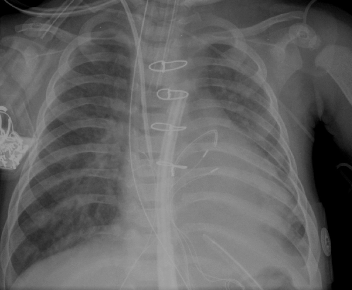
PA and Lateral views of venous and arterial catheter are provided.
Question 1: Trace the course of venous catheter. What diagnosis does this catheter-course leads you to?

Answer: Venous catheter-course: IVC - RA - PFO - LA - Left AV valve - morphologic RV (mRV) on the left side - Pulmonary valve - MPA - RPA. Diagnosis: Transposition of great arteries (L-TGA - Aortic valve is anterior in lateral view and to the left of the pulmonary valve in PA view)

Questoin 2: Trace the arterial catehter. Which side if the aortic arch?

Answer: Arterial catheter-course: Descending aorta - Aortic arch - Ascending aorta. Lateral view shows that the aorta is anterior. Aortic arch is left sided. Trachea is deviated towards the right. Apparent appearance of descending aorta on the right side of spine is misleading. Note the typical location of the ascending aorta in L-TGA. This causes a unique, fullness in the left, superior mediastinum in CXR.

Monday, June 11, 2012

Quiz 19: Answer

Echocardiogram: Aortic Arch View (Suprasternal notch view)

Question 1: What is abnormal in Panel 1?
Answer: (i) Left innominate vein is not see at the angle between the ascending aora and the origin of first arch vessel. (ii) Cross section of a second vessel is noted posterior to ascending aorta in addition to the usual right pulmonary artery.

Question 2: What vascular anomaly should be looked for?
Answer: Retroaortic innominate vein.
This is a rare entity noted in patient with Tetralogy of Fallot with Right aortic arch or when aorta has a "high" arch.

Clinical significance stems from rare clinical tales of the surgeon mistaking this retroaortic innominate vein for right pulmonary artery and places the BT shunt from subclavian artery to the innominate vein (& not to the right pulmonary artery).


Saturday, June 2, 2012

Quiz #18 Answer: EKG - AV Block, Second Degree

2-day old baby (Tetralogy of Fallot) developed this rhythm during cardiac catheterization.
Question: What is this catheter-induced rhythm?

Answer: Second degree AV Block

Arrows indicate P waves. There is 2:1 conduction. The conducted P waves are conducted with a prolonged PR interval (HR 83 bpm; PR interval 180-190 ms).

(At the onset, this was 3rd degree AV Block. This occurred during catheter manipulations in RV, attempting to cross a severely stenotic pulmonary valve. Intermittently, the catheter cross the VSD and entered the aorta. Presumably, the His bundle was affected during one of these times. Rhythm recovered spontaneously  to 2nd degree over time. Because the baby was unstable with ventricular rate of 83 bpm, Isoproterenol infusion was started after recording this EKG. Sinus rhythm was restrored after ~ 8 -12 hrs)

(Click on the image to enlarge)

Saturday, May 26, 2012

Quiz #17 Answer: Arterial line trace

Below is the image of ICU monitor of a teen ager.
Question 1: What is the name of the pulse pattern seen in the arterial line trace?
Answer: Pulsus paradoxus

Question 1: What are the 3 cause of such pulse pattern in any patient?
Answer: Pericardial tamponade, Severe asthma (Status asthmasticus) and Hypovolemia.

(Click on the image to enlarge)

Friday, May 18, 2012

Quiz #16: Answer (Echocardiogram - Pericardial effusion - M-mode)

Question: Image below is a parasternal short axis view recorded in a patient who presented with pericardial effusion. What ominous sign is being demonstrated?

Answer: Diastolic collapse of the RV free wall (Arrows. But, not seen in every beat due to movement artifacts). This is an echocardiographic sign of cardiac tamponade.

Tuesday, May 15, 2012

Quiz #15: 12-lead EKG - Inverted P in Lead I

Answer: There is P wave inversion in lead I and aVL. However, R wave progression in chest leads is normal. Therefore, this is misplacement of limb leads. Compare the corrected EKG when the same baby was 3 days old with normal lead placements.(Click on the images to enlarge)


Sunday, May 6, 2012

Quiz #14 Answer: EKG - Inverted P wave in lead I.


Question 1: What is the diagnosis?
Answer: Dextrocardia

Question 2: Provide 2 salient findings that support the diagnosis:
Answer: (i) Inverted P wave in leads I & aVL. (ii) Lack of R wave progression in chest leads (V1-V6)

Compare this EKG with that in Quiz #15.

Click on the image to enlarge.

Sunday, April 29, 2012

Quiz #13 Answer: Statistics - Z-score

A newborn (weight 3 kg, Length 50 cm) has aortic valve annulus of 6.0 mm. One published report states the following values for aortic valve annulus in newborns of the same body size: Mean 7.3 mm & Range of 6.38 – 8.22 mm. Range is reported as 2 standard deviations from the mean.
Question 1: What is the Z-score of the aortic valve in our patient with 6.0 mm diameter?
Answer: Z-score is (- 2.8)

Bonus Question: Please provide the steps of deriving the Z-score
Answer: Z-score is the represents the observed value as a the number of standard deviations away from the population mean. So, to calculate the Z-score for a given observed value, one has to know the population mean and SD.

Here the mean is 7.3 mm.
We need the SD. The given range is 2 SD on either sides of the mean. Therefore, 8.22 - 7.3 = 0.92 (which is equal to 2 SD).
SD = (0.92 ÷ 2) = 0.46

Given value for aortic valve diameter is 6.0 mm.
6.0 - mean = deviation from the mean.
i.e. 6.0 - 7.3 = (-1.3)

Deviation from mean ÷ SD = Z-score
i.e. (-1.3) ÷ 0.46 = (-2.8)
Z-score is (-2.8).

Thursday, April 12, 2012

Quiz #11 - Answer: CVP Trace

Question: The two snap shots were taken 1 minute apart from a postoperative patient.What is the difference between the CVP traces in each of the panels?

Answer: A white arrow has been added to each panel to help identify the "a" wave in each trace.
Top panel (Clock time 13:44 Hrs) - Recorded with A-pacing at 130 bpm. There is Atrio-ventricular synchrony. "a" wave is small (& is smaller than the "v" wave). Mean CVP is 7 mmHg. Arterial BP 92/46 (mean 64) mmHg.

Bottom panel (Clock time 13:45 Hrs) Again, the white arrow identifies the "a" wave. Comparatively, these are "giant" a waves. A-pacing is stopped. Patient's native rhythm is accelerated junctional rhythm at 104 bpm. AV synchrony is lost - atria are contracting against a closed AV valve causing "giant" a waves. Now, the CVP is 9 mmHg (higher than when there was AV synchrony) and arterial BP is lower (73/33, mean 46 mmHg) - emphasizing the importance of AV synchrony in this patient.

Note: "Cannon" a waves are occasional giant a waves seen in patients with complete AV block. In this situation, a waves are normal or close to normal during several beats when there is apparent AV synchrony by shear coincidence of a sequential atrial and ventricular contractions. But, there will be occasional sudden appearance of one giant a wave at variable intervals when apparent AV synchrony is lost.


Sunday, April 8, 2012

Quiz 10 Answer: Clinical Examination

Answers:
What is the sign shown in figure 1?
Answer: Wrist sign

What is the sign shown in figure 2?
Answer: Thumb sign

If a patient has both signs, he/she gets 3 points in the "systems score" - according to the Revised Ghent Criteria for diagnosis of Marfan syndrome.
(Click here for System Score table)

Other links for information about Marfan syndrome:
http://www.ncbi.nlm.nih.gov/books/NBK1335/#marfan.Diagnosis
http://www.marfan.org/marfan/4265/Diagnostic-Criteria


Saturday, March 31, 2012

Quiz #9: Where is the tip of the PA line?

Question: Based on the CXR and PA line pressure trace, what is the location of the PA line tip? (Clue: The PA pressure trace holds the answer. Chest Xray helps to corroborate.)

Answer: RV outflow tract.

Rationale: PA pressure has a ventricular trace with a pressure of 55/1. If you enlarge and see, pressure trace is typical of a ventricular pattern. So, the line tip is in the RV. But, in the CXR, it is high up in the RV. Combining the two findings, the line tip has to be below the valve i.e. RVOT.

Of course, this patient has elevated PA pressure at ~50% systemic level.


Friday, March 23, 2012

Quiz #8: Barium Swallow

Question 1: What are the salient findings on Barium Swallow?
Answer 1: Bilateral indentation of the esophagus.
(Indentation on the right side is at a higher level than the left side which is usual in this condition)


Question 2: What is the diagnosis?
Answer 2: Double aortic arch (MRI from the same patient is given below).

Saturday, March 17, 2012

Quiz #7: Barium Swallow Study


Question 1: Describe the findings in PA view?

Answer 1: Oblique impression in the upper esophagus (going left to right, from below upwards). However, in addition, there is a subtle compression of both side of the trachea.


Question 2: Describe the finding in Lateral view?

Answer 2: Posterior indentation of esophagus in the upper esophagus.


Question 3: What is the diagnosis?

Answer 3: Anomalous origin of right subclavian artery. In addition, this baby also has a possibility of double aortic arch (with patent right side arch giving off all the branches and ligamentous representation of left side arch. Left side arch is a presumption based on the the following findings: Right aortic arch with left descending aorta - associated with a Kommerell's diverticulum). See the rule in "Moss & Adams" - Vascular ring chapter by Dr. Paul Weinberg. Click here for link to a blog posting about the "rule". Briefly, the rule states that presence of 3 Ds in the opposite side from the ascending aorta, there is a vascular ring. The 3 Ds are Diverticulum, Dimple or Descending aorta.


Saturday, March 10, 2012

Quiz #6: Barium Swallow Answers

Link to the question

Question 1: What is salient finding?
Answer: Anterior indentation of the esophagus. This means, the structure is passing inbetween the trachea and esophagus. The indentation in this condition is usually at the level of carina.

Question 2: What is the diagnosis?
Answer: "Pulmonary sling". This term refers to anomalous origin of LPA from RPA. The LPA courses inbetween trachea and esophagus to reach the left side.

2 bonus questions:
Question 3: What is the tracheal anomaly associated with pulmonary sling?
Answer: Complete rings in distal trachea - that may need additional surgical treatment (apart from correcting vascular anomaly)

Question 4: What cardiac anomaly is commonly associated with pulmonary sling?
Answer: Usually occurs by itself. Common associated cardiac lesion is Tetralogy of Fallot.
(Ref: Moss & Adams Fifth edition. p. 833. Chapter by Dr. Paul Weinberg)

7 out of 7 fellows responded. One fellow needed some "proding". But, ultimately everyone got the answer right. Dr. Agu sent a great article on this subject (Thorax 1969;24:295-306) with very illustrative figures.

The 2 MRI images are from one of our patients (Black arrow - Trachea. White arrow - Esophagus with NG tube). This patient did not have a barium swallow. Such is the trend these days! Barium swallow is still a good, straight forward study to demonstrate esophageal compression. Sometimes, this is unclear in MRI images.


Sunday, February 26, 2012

Quiz #5 Answers: WPW syndrome

Click on the image to enlarge.
Questions:
1) What is the diagnosis?
2) Where is the pathway located?


Answers:
1) WPW syndrome (Short PR interval and Delta wave)
2) Right posteroseptal accessory pathway

Thanks to Dr. Numan for advice with interpretation.

There are several algorithms. Two are given below. None of them is perfect. But, they help.
(Click on the image to see a large image)
Bibliography:
Fitzpatrick et al. J Am Coll Cardiol. 1994 Jan;23(1):107-16, (Erratum in
J Am Coll Cardiol 1994 Apr;23(5):1272).
Fitzpatrick J Electrocardiol. 1993;26 Suppl:220-6.
Fitzpatrick PACE 1995.
Arruda et al. J Cardiovasc Electrophysiol 1998;9:2-12.
http://awolecg.blogspot.com/2009/10/wpw-localization-of-accessory-pathway.html
& there are many more!


Monday, February 20, 2012

Answer to Quiz #4: EKG - Pacemaker

Question: What's wrong?


Answer:

Ventricular wire is inserted into atrial outlet of the pacemaker and atrial wire into the ventricular outlet!


Finding in the EKG strip: In each beat, first pacing-spike elicits a QRS complex followed by the second pacing spike that elicits a P wave!

Friday, February 10, 2012

Answer to Quiz #3: Surgical anatomy - Arrangement of Atrial Appendages

Answer:
Arrangement shown is juxtaposition of atrial appendages. First figure is juxtapositioning of atrial appendages to the right side of great arteries. Second figure is juxtapositioning of atrial appendages to the left side of great arteries.

Left juxtapositioning (86%) is more common than right juxtaposition (14%) - based on autopsy study of 49 cases with juxtaposition.

Left juxtaposition is associated with complex heart defects more often than right juxtaposition.
Common associations with left juxtaposition are (i) Tricuspid atresia, (ii) Transposition and (iii) DORV.
Common associations with right juxtaposition are (i) Outflow tract obstruction, (ii) Anomalous venous return and (iii) DILV.

Ref:
1) Echocardiography in Pediatric Heart Disease by R. Snider et al. 2nd ed. 1997 (p.562)
2) Cardiovascular Pathology 2011; published online Apr 11, 2011


Saturday, February 4, 2012

Answer to Quiz #2: Scimitar Syndrome

Answer:

Abdominal aortogram shows accessory pulmonary blood flow via aorto-pulmonary collateral arteries. The pulmonary venous return (at the end of the angiogram) returns to IVC-RA junction constituting partial anomalous pulmonary venous return.

This is scimitar syndrome.

Scimitar syndrome is characterized by (i) Lung sequestration (usually, right lower lobe), (ii) Accessory blood flow to the sequestered segment from abdominal aorta and (iii) Partial anomalous pulmonary venous return - drainage to IVC).


Pulmonary hypertension may occur in newborn and infants. Exact pathogenesis of pulmonary hypertension in Scimitar syndrome is unknown. Neonatal or infantile presentation with symptoms and presence of pulmonary hypertension is a bad prognostic signs.


Recurrent pneumonia in the sequestered lung segment is an indication for removal of the sequestered lung segment.


Transcatheter intervention: Coil occlusion of acc. pulmonary blood flow helps to reduce the vascularity. When the surgeon goes to remove the sequestered lung segment, prior coil occlusion of accessory blood flow to the sequestered segment saves the surgeon having to deal with ligating the blood vessels in the abdomen.


Journal article on Scimitar syndrome - 20 year experience from Toronto.


Monday, January 30, 2012

Answer to Quiz #1: What is the surgery (surgeries) shown in this image?


Answer:
Components of the surgery shown in the image are the following:
1) Classic/Unidirectional Glenn anastamosis (SVC - distal RPA connection)
2) RA connected to proximal RPA stump.
3) PA band or PA ligation
4) Two biological valve: one between IVC and RA, other between RA and RPA. This was supposed to ensure that RA contractility - thought to be important as a pump for pulmonary circulation - can be transmitted only forward, into pulmonary circulation.
5) ? Right atrial appendage anastamosed to RVOT (Not sure if this was part of original Fontan operation - published in 1971.

As we know, much of this operation has changed since 1971. But, the concept remains the only solution for children with single ventricle.

Differences between 1971 Classic Fontan and the figure in WJPCHS are the following:


1) In Classic Fontan, a pulmonary homograft valve was placed inside the native IVC-RA junction (In 2012 image, it appears the there is interpositioning of a piece of homograft. This would entail disconnecting IVC from RA and then, placing this homograft.


2) In Classic Fontan, an aortic homograft was sutured to the proximal stump of RPA first. Then, the other end of the aortic homograft was sutured to RA appendage. (In 2012 image, RA appendage seems to remain undisturbed. the aortic homograft is sutured on to top of the right atrial wall.

(Picture of "Classic" Fontan operation is from Fontan, F. & Baudet, E. Surgical Repair of Tricuspid Atresia. Thorax 1971;26:240-248)