CT scan images from a 8-mo old boy with stridor, vomiting
and weighs 5.6 kg. Figure 1 shows anterior and Left lateral views. Figure 2
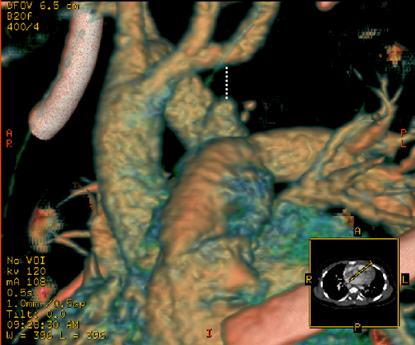
shows 3D reconstruction of trachea-bronchial tree (Barium swallow was not
performed in this patient).
Questions:
1) What is the arch sidedness?
2) What is the diagnosis?
3) Does the patient need intervention and why?
Answers:
1) Right aortic arch (Arrow in the last image shows indentation in trachea).
2) Double arch with ligamentous (atretic), left arch - creating a vascular ring.
3) Patient needs intervention because the patient is symptomatic and failing to thrive.
A differential interpretation for the diagnosis - Question 2 - is "Right aortic arch with left ductal ligament". Where the anterior end of the ligament is attached will determine the difference. If the ligament is attached to the ascending aorta or a branch of the aorta, it is "double arch". Alternatively, if the ligament is attached to the MPA, it is "right arch with ductal ligament". Dotted line is drawn in the images below to help to make this determination.
Arrow in the last figure indicates the indentation in trachea from right-sided arch. Similar indentation is expected in barium swallow.


No comments:
Post a Comment
Comments are moderated.