Friday, May 18, 2012

Quiz #16: Answer (Echocardiogram - Pericardial effusion - M-mode)

Question: Image below is a parasternal short axis view recorded in a patient who presented with pericardial effusion. What ominous sign is being demonstrated?

Answer: Diastolic collapse of the RV free wall (Arrows. But, not seen in every beat due to movement artifacts). This is an echocardiographic sign of cardiac tamponade.

Tuesday, May 15, 2012

Quiz #15: 12-lead EKG - Inverted P in Lead I

Answer: There is P wave inversion in lead I and aVL. However, R wave progression in chest leads is normal. Therefore, this is misplacement of limb leads. Compare the corrected EKG when the same baby was 3 days old with normal lead placements.(Click on the images to enlarge)


Sunday, May 6, 2012

Quiz #14 Answer: EKG - Inverted P wave in lead I.


Question 1: What is the diagnosis?
Answer: Dextrocardia

Question 2: Provide 2 salient findings that support the diagnosis:
Answer: (i) Inverted P wave in leads I & aVL. (ii) Lack of R wave progression in chest leads (V1-V6)

Compare this EKG with that in Quiz #15.

Click on the image to enlarge.

Sunday, April 29, 2012

Quiz #13 Answer: Statistics - Z-score

A newborn (weight 3 kg, Length 50 cm) has aortic valve annulus of 6.0 mm. One published report states the following values for aortic valve annulus in newborns of the same body size: Mean 7.3 mm & Range of 6.38 – 8.22 mm. Range is reported as 2 standard deviations from the mean.
Question 1: What is the Z-score of the aortic valve in our patient with 6.0 mm diameter?
Answer: Z-score is (- 2.8)

Bonus Question: Please provide the steps of deriving the Z-score
Answer: Z-score is the represents the observed value as a the number of standard deviations away from the population mean. So, to calculate the Z-score for a given observed value, one has to know the population mean and SD.

Here the mean is 7.3 mm.
We need the SD. The given range is 2 SD on either sides of the mean. Therefore, 8.22 - 7.3 = 0.92 (which is equal to 2 SD).
SD = (0.92 ÷ 2) = 0.46

Given value for aortic valve diameter is 6.0 mm.
6.0 - mean = deviation from the mean.
i.e. 6.0 - 7.3 = (-1.3)

Deviation from mean ÷ SD = Z-score
i.e. (-1.3) ÷ 0.46 = (-2.8)
Z-score is (-2.8).

Thursday, April 12, 2012

Quiz #11 - Answer: CVP Trace

Question: The two snap shots were taken 1 minute apart from a postoperative patient.What is the difference between the CVP traces in each of the panels?

Answer: A white arrow has been added to each panel to help identify the "a" wave in each trace.
Top panel (Clock time 13:44 Hrs) - Recorded with A-pacing at 130 bpm. There is Atrio-ventricular synchrony. "a" wave is small (& is smaller than the "v" wave). Mean CVP is 7 mmHg. Arterial BP 92/46 (mean 64) mmHg.

Bottom panel (Clock time 13:45 Hrs) Again, the white arrow identifies the "a" wave. Comparatively, these are "giant" a waves. A-pacing is stopped. Patient's native rhythm is accelerated junctional rhythm at 104 bpm. AV synchrony is lost - atria are contracting against a closed AV valve causing "giant" a waves. Now, the CVP is 9 mmHg (higher than when there was AV synchrony) and arterial BP is lower (73/33, mean 46 mmHg) - emphasizing the importance of AV synchrony in this patient.

Note: "Cannon" a waves are occasional giant a waves seen in patients with complete AV block. In this situation, a waves are normal or close to normal during several beats when there is apparent AV synchrony by shear coincidence of a sequential atrial and ventricular contractions. But, there will be occasional sudden appearance of one giant a wave at variable intervals when apparent AV synchrony is lost.


Sunday, April 8, 2012

Quiz 10 Answer: Clinical Examination

Answers:
What is the sign shown in figure 1?
Answer: Wrist sign

What is the sign shown in figure 2?
Answer: Thumb sign

If a patient has both signs, he/she gets 3 points in the "systems score" - according to the Revised Ghent Criteria for diagnosis of Marfan syndrome.
(Click here for System Score table)

Other links for information about Marfan syndrome:
http://www.ncbi.nlm.nih.gov/books/NBK1335/#marfan.Diagnosis
http://www.marfan.org/marfan/4265/Diagnostic-Criteria


Saturday, March 31, 2012

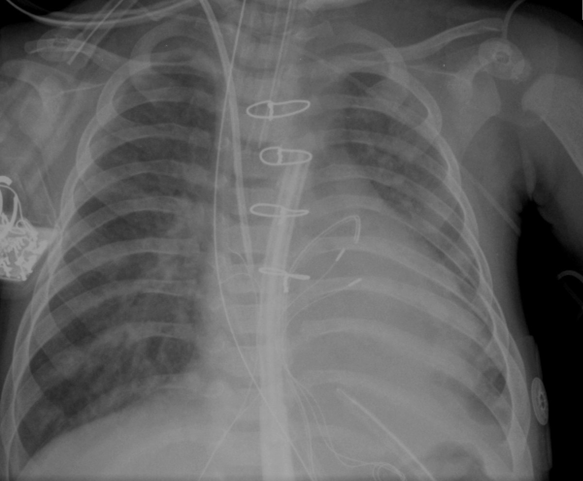
Quiz #9: Where is the tip of the PA line?

Question: Based on the CXR and PA line pressure trace, what is the location of the PA line tip? (Clue: The PA pressure trace holds the answer. Chest Xray helps to corroborate.)

Answer: RV outflow tract.

Rationale: PA pressure has a ventricular trace with a pressure of 55/1. If you enlarge and see, pressure trace is typical of a ventricular pattern. So, the line tip is in the RV. But, in the CXR, it is high up in the RV. Combining the two findings, the line tip has to be below the valve i.e. RVOT.

Of course, this patient has elevated PA pressure at ~50% systemic level.