Friday, March 23, 2012

Quiz #8: Barium Swallow

Question 1: What are the salient findings on Barium Swallow?
Answer 1: Bilateral indentation of the esophagus.
(Indentation on the right side is at a higher level than the left side which is usual in this condition)


Question 2: What is the diagnosis?
Answer 2: Double aortic arch (MRI from the same patient is given below).

Saturday, March 17, 2012

Quiz #7: Barium Swallow Study


Question 1: Describe the findings in PA view?

Answer 1: Oblique impression in the upper esophagus (going left to right, from below upwards). However, in addition, there is a subtle compression of both side of the trachea.


Question 2: Describe the finding in Lateral view?

Answer 2: Posterior indentation of esophagus in the upper esophagus.


Question 3: What is the diagnosis?

Answer 3: Anomalous origin of right subclavian artery. In addition, this baby also has a possibility of double aortic arch (with patent right side arch giving off all the branches and ligamentous representation of left side arch. Left side arch is a presumption based on the the following findings: Right aortic arch with left descending aorta - associated with a Kommerell's diverticulum). See the rule in "Moss & Adams" - Vascular ring chapter by Dr. Paul Weinberg. Click here for link to a blog posting about the "rule". Briefly, the rule states that presence of 3 Ds in the opposite side from the ascending aorta, there is a vascular ring. The 3 Ds are Diverticulum, Dimple or Descending aorta.


Saturday, March 10, 2012

Quiz #6: Barium Swallow Answers

Link to the question

Question 1: What is salient finding?
Answer: Anterior indentation of the esophagus. This means, the structure is passing inbetween the trachea and esophagus. The indentation in this condition is usually at the level of carina.

Question 2: What is the diagnosis?
Answer: "Pulmonary sling". This term refers to anomalous origin of LPA from RPA. The LPA courses inbetween trachea and esophagus to reach the left side.

2 bonus questions:
Question 3: What is the tracheal anomaly associated with pulmonary sling?
Answer: Complete rings in distal trachea - that may need additional surgical treatment (apart from correcting vascular anomaly)

Question 4: What cardiac anomaly is commonly associated with pulmonary sling?
Answer: Usually occurs by itself. Common associated cardiac lesion is Tetralogy of Fallot.
(Ref: Moss & Adams Fifth edition. p. 833. Chapter by Dr. Paul Weinberg)

7 out of 7 fellows responded. One fellow needed some "proding". But, ultimately everyone got the answer right. Dr. Agu sent a great article on this subject (Thorax 1969;24:295-306) with very illustrative figures.

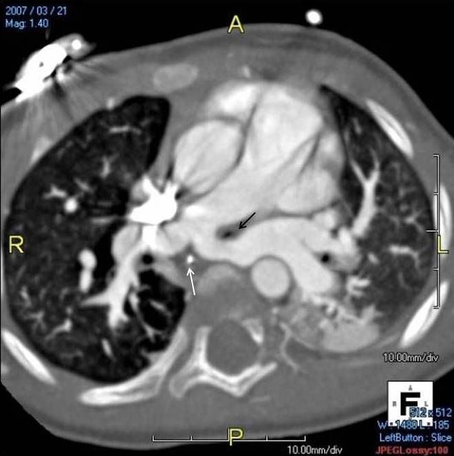
The 2 MRI images are from one of our patients (Black arrow - Trachea. White arrow - Esophagus with NG tube). This patient did not have a barium swallow. Such is the trend these days! Barium swallow is still a good, straight forward study to demonstrate esophageal compression. Sometimes, this is unclear in MRI images.




Sunday, February 26, 2012

Quiz #5 Answers: WPW syndrome

Click on the image to enlarge.
Questions:
1) What is the diagnosis?
2) Where is the pathway located?


Answers:
1) WPW syndrome (Short PR interval and Delta wave)
2) Right posteroseptal accessory pathway

Thanks to Dr. Numan for advice with interpretation.

There are several algorithms. Two are given below. None of them is perfect. But, they help.
(Click on the image to see a large image)
Bibliography:
Fitzpatrick et al. J Am Coll Cardiol. 1994 Jan;23(1):107-16, (Erratum in
J Am Coll Cardiol 1994 Apr;23(5):1272).
Fitzpatrick J Electrocardiol. 1993;26 Suppl:220-6.
Fitzpatrick PACE 1995.
Arruda et al. J Cardiovasc Electrophysiol 1998;9:2-12.
http://awolecg.blogspot.com/2009/10/wpw-localization-of-accessory-pathway.html
& there are many more!


Monday, February 20, 2012

Answer to Quiz #4: EKG - Pacemaker

Question: What's wrong?


Answer:

Ventricular wire is inserted into atrial outlet of the pacemaker and atrial wire into the ventricular outlet!


Finding in the EKG strip: In each beat, first pacing-spike elicits a QRS complex followed by the second pacing spike that elicits a P wave!

Friday, February 10, 2012

Answer to Quiz #3: Surgical anatomy - Arrangement of Atrial Appendages

Answer:
Arrangement shown is juxtaposition of atrial appendages. First figure is juxtapositioning of atrial appendages to the right side of great arteries. Second figure is juxtapositioning of atrial appendages to the left side of great arteries.

Left juxtapositioning (86%) is more common than right juxtaposition (14%) - based on autopsy study of 49 cases with juxtaposition.

Left juxtaposition is associated with complex heart defects more often than right juxtaposition.
Common associations with left juxtaposition are (i) Tricuspid atresia, (ii) Transposition and (iii) DORV.
Common associations with right juxtaposition are (i) Outflow tract obstruction, (ii) Anomalous venous return and (iii) DILV.

Ref:
1) Echocardiography in Pediatric Heart Disease by R. Snider et al. 2nd ed. 1997 (p.562)
2) Cardiovascular Pathology 2011; published online Apr 11, 2011


Saturday, February 4, 2012

Answer to Quiz #2: Scimitar Syndrome

Answer:

Abdominal aortogram shows accessory pulmonary blood flow via aorto-pulmonary collateral arteries. The pulmonary venous return (at the end of the angiogram) returns to IVC-RA junction constituting partial anomalous pulmonary venous return.

This is scimitar syndrome.

Scimitar syndrome is characterized by (i) Lung sequestration (usually, right lower lobe), (ii) Accessory blood flow to the sequestered segment from abdominal aorta and (iii) Partial anomalous pulmonary venous return - drainage to IVC).


Pulmonary hypertension may occur in newborn and infants. Exact pathogenesis of pulmonary hypertension in Scimitar syndrome is unknown. Neonatal or infantile presentation with symptoms and presence of pulmonary hypertension is a bad prognostic signs.


Recurrent pneumonia in the sequestered lung segment is an indication for removal of the sequestered lung segment.


Transcatheter intervention: Coil occlusion of acc. pulmonary blood flow helps to reduce the vascularity. When the surgeon goes to remove the sequestered lung segment, prior coil occlusion of accessory blood flow to the sequestered segment saves the surgeon having to deal with ligating the blood vessels in the abdomen.


Journal article on Scimitar syndrome - 20 year experience from Toronto.